
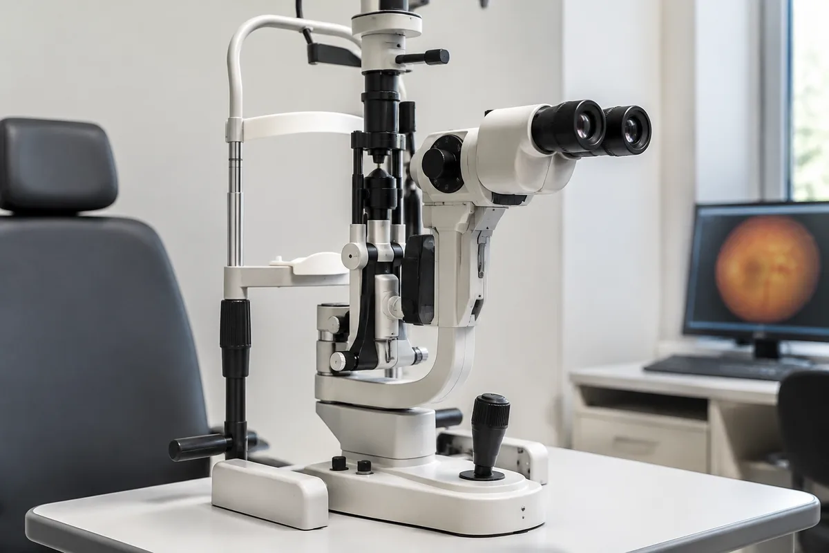
A slit lamp microscope (also called a biomicroscope) is one of the most important instruments in a comprehensive eye exam. It pairs a binocular microscope with a bright, adjustable beam of light so your provider can see the structures at the front of your eye in fine, magnified detail.
How It Works
The instrument projects a narrow "slit" of light that can be widened, narrowed, angled and filtered. Viewed through the microscope, that beam lights up the eyelids, lashes, conjunctiva, cornea, iris and lens in three dimensions, and with special handheld lenses it can also be used to look at the retina and optic nerve at the back of the eye.
Why It's Used
A slit lamp exam helps detect and monitor a wide range of conditions, including dry eye, corneal scratches and infections, inflammation, cataracts, and signs of glaucoma or macular degeneration. It is also used to check the fit of contact lenses and to follow healing after an injury or eye surgery.
What to Expect
You'll rest your chin and forehead on the instrument's supports while your provider looks through the microscope. The exam is painless and only takes a few minutes; depending on what is being checked, eye drops may be used to dilate your pupils or a harmless yellow dye may be applied to highlight the surface of the cornea.
This content is for general information only and isn't medical advice. Talk with a qualified provider about your own situation. If you have a medical emergency, call 911.